


The ankle joint is often injured due to the heavy load it is subjected to. A diagnosis such as arthrosis of the ankle is not uncommon. It is placed regardless of the patient's age and gender. What is ankle osteoarthritis and how can it be treated?
What is it?
There is a huge load on the ankle. Its function is to keep the body erect. Thanks to him, a person walks and runs. With a breach of the ankle system, it is extremely difficult to lead a family lifestyle. What hinders the ankle work?
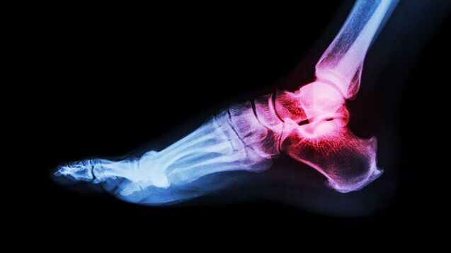
Arthrosis of the ankle, what is it? It is a chronic disease of the joints, characterized by a degenerative course. In the joint cartilage, irreversible processes are unleashed, which lead to formidable complications.
Ankle arthrosis develops gradually. Healthy joint surfaces are elastic and smooth. They provide cushioning under heavy loads and smooth glide while driving. With the pathology, trophism and tissue metabolism are disturbed. The joint surface becomes inelastic and rough. During movement, the cartilages come into contact with each other, which leads to inflammation. When lifting weights, the main load falls on the bone, which threatens degenerative diseases.
Lack of treatment leads to more serious disorders. In 3-4 stages, cartilage and tissue damage is seen. The synovium becomes inflamed. The joint becomes unstable. The support function has been violated. All these violations together lead to the fact that movement becomes impossible.
Arthrosis (osteoarthritis) is one of the most common joint diseases that affects a very large number of people.
Causes and risk factors
What is arthrosis of the ankle joint, we've solved it. Now let's find out what its root cause is. Ankle arthrosis is considered a condition of old age. This is due to age-related changes in the body. Cartilage becomes thinner, bones become unstable and fragile. However, in the last decade, the diagnosis of ankle arthrosis has become much younger. These statistics are disappointing, as many patients ignore the first signs of the disease. Late diagnosis always threatens the development of serious complications.
Provoking factors include:
- displacements;
- bruises;
- inflammatory diseases;
- prejudice;
- overweight;
- impaired metabolism;
- unbearable physical activity;
- wear uncomfortable shoes;
- autoimmune and endocrine diseases;
- osteochondrosis.
clinical symptoms
Ankle arthrosis is recognized by the following characteristics:
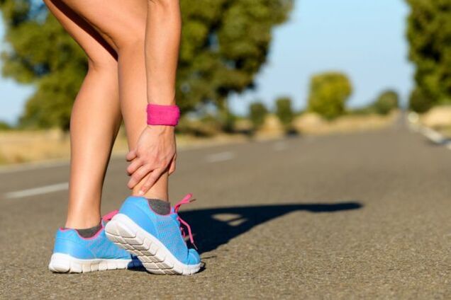
- Pain. It is light at first and appears after walking or physical exertion. Sometimes when a person is in an uncomfortable position. With the progression of the pathological process, the painful syndrome intensifies and worries already at rest.
- Swelling and inflammation. These signs appear in the context of injuries and dislocations. The body temperature in the affected area increases.
- Click. When the ankle is affected, the click becomes "dry" and causes an attack of pain.
- Dislocation or subluxation. Due to the thinning and degradation of cartilage tissue, the joint becomes unstable. Bones can shift and fall out of the joint capsule. These changes cause acute pain crises.
- Joint stiffness. When cartilage tissue is replaced, the bone joint ceases to function normally, which negatively affects its mobility.
- Joint deformity. The symptom appears in 3-4 stages of arthrosis. Osteophytes also cause ankle curvature.
If one of the symptoms appears, it is recommended to see a doctor immediately. Treatment started in a timely manner is a step towards recovery.
Arthrosis of the foot and ankle joints is characterized by a slow progression with a gradual development of clinical manifestations over several years.
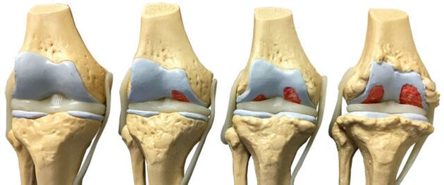
classification and stages
The disease develops in different ways. In some patients, several years pass from the first signs to the final stage, in others, the rapid development of the disease is observed. The speed depends on the age and state of health of the patient, when the therapy starts. The symptoms of arthrosis of the ankle joint become more intense as the disease progresses.
There are four stages of arthrosis:
- The first stage often goes unnoticed. Sometimes morning stiffness and ankle pain appear after heavy exertion. When the foot moves, there is a characteristic click. Pathological changes are not yet visible on radiographs, but the destructive process of the cartilage has already begun.
- Morning stiffness becomes prolonged. It takes 20-30 minutes to develop a leg. Sometimes lameness occurs. Second-degree arthrosis of the ankle joint is recognized on radiography by the growth of bone tissue, displacement of the bones.
- 3-stage symptoms are pronounced. Pain is no longer just worrying after a heavy load, but also at rest. It is difficult for a patient to live without painkillers. The lameness increases. Crutches may be needed. The affected joint is swollen and deformed. Ankle muscles atrophy. X-ray shows joint space narrowing, osteophyte formation, subluxation.
- Stage 4 is the hardest. It develops as a result of lack of treatment. Cartilage is destroyed, joint surfaces fuse. Walking is no longer possible.
With the development of ankle osteoarthritis, there is a gradual change in the cartilage and bone tissue of the joint surfaces.
Diagnosis
The diagnosis of ankle arthrosis is based on clinical symptoms and information obtained during examinations. Laboratory studies are considered ineffective, as there are no special tests that can detect the pathology. During the remission period, all indicators are within normal limits, with an exacerbation of the disease, a clinical blood test will show a high level of C-reactive protein and ESR. These indicators indicate that the pathological process has already started.
To confirm the diagnosis, instrumental methods are used:
- radiography;
- Magnetic resonance imaging;
- Ultrasound;
- bone scintigraphy;
- diagnostic joint puncture.
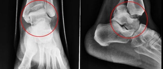
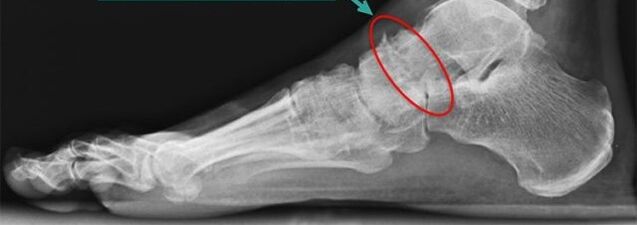
plain radiography
Plain radiography is the most reliable and effective method for diagnosing diseases that occur in the musculoskeletal system. The principle of manipulation is the differentiated absorption of X-rays by muscle tissue. Soft tissues allow X-rays to pass through, but hard tissues are absorbed. An X-ray allows you to diagnose the disease itself and its consequences.
Conventional radiography is an examination method in which a small amount of X-rays are transmitted through the person's body or part of the body.
The snapshot lets you see:
- The condition of bone surfaces in the joint.
- The shape, size and arrangement of the structures in the joint are relative to each other.
- The state of the tissue.
- The size of the joint space.
These indicators help the doctor determine the type and extent of joint damage. If the data are not enough, doctors prescribe further studies.
With ankle arthrosis, an X-ray is taken in three projections:
- side;
- back;
- back with one foot moved inward.
The disease is characterized by the following changes:
- joint space reduction;
- the presence of osteophytes;
- bone cartilage replacement (subchondral sclerosis);
- small voids in the periarticular part.
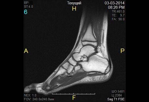
nuclear magnetic resonance
Nuclear magnetic resonance (NMR) as a diagnostic method makes it possible to study the parts of the body where there is water. The image shows bones in darker colors as they contain less water, but muscle tissue, discs and nerves are shown lighter. Magnetic resonance allows detecting the smallest changes in the structure of bone tissue and joints. The study is also prescribed for patients before joint prosthesis. YMG has a downside - a high price.
In nuclear magnetic resonance, a change in the properties of hydrogen molecules under the influence of a strong magnetic field is recorded.
magnetic resonance imaging
Magnetic resonance imaging (MRI) is an alternative diagnostic method that allows you to carefully examine the ligamentous structure of the joint, muscle, and cartilage tissue. With the help of an MRI, the doctor assesses the condition of the leg joints. Based on research data, the pathology is revealed at an early stage of development.
The diagnostic principle is based on exposure to radio waves and strong magnetic radiation. The magnetic field used is not hazardous and does not pose a health hazard.
Magnetic resonance imaging is contraindicated in case of mental disorders, during pregnancy and in the presence of metal objects in the human body.
In the diagnosis of ankle arthrosis, classical magnetic resonance machines (closed type) are used, as they have better image quality. An MRI machine is a large cylindrical tube with a magnet around it. The patient lies down on a special table. The ankle is secured with a special coil. The procedure takes 30 to 40 minutes. The study is absolutely painless. Patients may feel heat in the leg region.
ultrasound
Ultrasound examination has been widely used in medicine since the 90's of the 20th century. This technique has shown itself well for making accurate diagnoses. An ultrasound is also performed for ankle joint arthrosis.
Today, the ultrasound examination is not particularly important in the diagnosis of osteoarthritis, as it does not allow for a good enough study of injured joints.
The device with which the study is carried out produces ultra-frequency waves. Waves are reflected from the tissues and recorded on the monitor. Based on the resulting image, the doctor determines the type of pathology. To make the image sharp on the monitor, a special gel is used. Eliminates air gaps and gives the sensor a better glide.
The ultrasound exam does not harm the patient, so the procedure can be repeated several times. The advantages of ultrasound also include low cost and high accuracy.
The following indicators are a clear sign of osteoarthritis:
- cartilage thinning;
- the presence of bone growths;
- accumulation of effusion in the joint cavity (synovitis);
- loss of cartilage space.
bone scintigraphy
Scintigraphy is a high-precision study that, through isotopes, is capable of detecting pathological changes in bones. Doctors divide pathogenic foci into "cold" and "hot". In the first case, we are talking about zones where there are no isotopes. These areas are poorly supplied with blood and are not visible during the examination. "Cold" areas are places affected by malignant tumors. In "hot" areas, isotopes build up quickly and look very bright when scanned. These areas indicate the presence of inflammatory processes.
The role of scintigraphy in arthrosis is significant. The study helps to distinguish osteoarthritis from several other diseases when clinical symptoms are extremely similar.
During bone scintigraphy, a special preparation containing special marked atoms is injected into the body.
Based on the results of the scintigraphy, the physician makes a clinical prognosis and determines the treatment regimen. The only disadvantage of the study is its high cost. Scintigraphy is performed with special equipment and unfortunately not all medical institutions can afford it.
Although radioactive scanning is a safe procedure, it still has a number of contraindications:
- pregnancy;
- lactation period;
- taking medications containing barium.
When a radioactive substance is injected, some patients experience an allergic reaction in the form of itching and rash. These side effects are harmless and go away on their own in a short time.
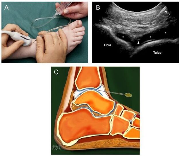
joint puncture
Joint puncture is a diagnostic procedure in which a needle is inserted into the joint cavity to collect synovial fluid. This liquid is then sent for further research. Based on the data obtained, the physician concludes about the nature of the disease and the stage of its development.
At first glance, puncture is a simple procedure, but it is not. The removal of fluid from the joint capsule requires exceptional precision of the physician's movements. The synovium is very thin and a strange movement traumatizes it. As a result, an inflammatory process develops. Potential risks also include infection. It is not difficult to bring the infection to the joint capsule through poorly sterilized instruments.
The manipulation technique is different for each joint. When collecting the ankle joint exudate, the puncture is performed in front, between the external ankle and the tendon of the extensor digitorum longus.
Diagnostic sampling of intra-articular fluid allows for laboratory analysis and excludes inflammatory arthritis.
Basic treatment principles
After confirming the diagnosis of arthrosis of the ankle joint, symptoms will soon appear. Treatment starts immediately. Additional prognosis depends on a well-chosen treatment regimen and timing of initiation.
Arthrosis is an insidious disease. It cannot be completely cured. The aim of therapy is to stop the degenerative processes and prolong the remission period. To do so, doctors prescribe medication, physiotherapy, massage, corrective gymnastics and folk remedies. If all conditions are met, it is possible to count on a positive dynamic, otherwise the disease progresses.
Drug therapy for arthrosis
Depending on the therapeutic effect, drugs are divided into several groups:
- Anti-inflammatories or analgesics. This group of medications aims to eliminate the focus of inflammation and relieve pain. The earlier anti-inflammatory therapy is started, the greater the chances of saving the joint. Medicines in this group can be produced in the form of pills and ointments.
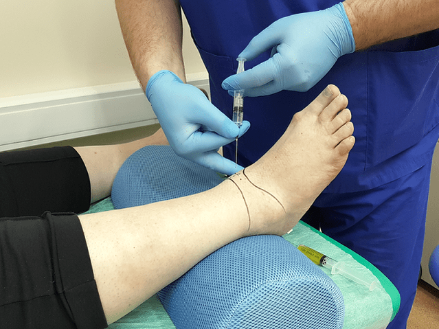
- Glucocorticoids. These medications are prescribed when the above funds are ineffective. They are produced in the form of an injectable solution. The drug is injected directly into the joint.
- Chondroprotective. Designed to slow cartilage destruction.
The treatment regimen and dosage are selected by the physician based on the severity of symptoms, the age of the patient, the presence of concomitant illnesses, and other factors. Self-medication is dangerous and often makes the situation worse, as many medications have various side effects and have their own contraindications.

Characteristics of radical treatment
If conservative therapy has failed, doctors are forced to resort to a radical method of treatment (surgical intervention). The operation is also shown when:
- secondary (posttraumatic) and primary arthrosis of 3-4 degrees;
- arthrosis with complications;
- constant severe pain in the ankle, radiating to the knee;
- severe lameness;
- paresis and paralysis of leg muscles;
- violation of the flexo-extensor function of the joint;
- violation of the foot's ability to support.
Surgical intervention is contraindicated if:
- the patient is less than 12 years old;
- fistulas are found in the joint;
- the patient has a history of diabetes mellitus, heart failure;
- Infectious diseases were found in the proposed intervention area.
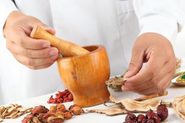
traditional treatment
Doctors believe that the treatment of osteoarthritis should be carried out exclusively under the supervision of a specialist, but they do not deny the positive effect of folk remedies. Alternative medicine acts as an effective prophylaxis that helps eliminate symptoms and maintain remission.
Folk remedies are a very symptomatic treatment for foot arthrosis.
Home treatment should be coordinated with your doctor to avoid side effects and complications.
Traditional healers suggest treating ankle arthrosis with:
- Burdock. Wash burdock leaves with soap and running water. Apply the leaves with the soft side to the skin. Secure the top with a bandage or plastic wrap. It's best to keep the compress overnight.
- Sea salt. Chop the salt in a skillet. Pour it into a linen bag and secure it to your ankle. Hold the bag until the salt cools. Heat relieves pain. Sand, lentils, buckwheat are also used instead of salt.
- Lilacs. Pour the triple colony over the lavender flowers. Let the dye rest in a cool, dark place for 10-14 days. Rub the affected area morning and night.
- Eggshell. Grind the husks in a coffee grinder. Take the resulting powder for ½ tsp. before eating.
Don't forget that treatment with folk remedies should not be the only measure. Complex treatment includes taking medication, exercise therapy, massage, physiotherapy, spa treatment. In advanced cases, doctors resort to radical measures - surgical intervention.
Surgery
For foot arthrosis, the following types of operations are used in medicine:
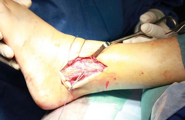
- joint arthrodesis;
- joint arthroscopy;
- endoprosthesis.
Arthrodesis is an operation to immobilize a joint. It is carried out with the aim of returning the member to the lost carrying capacity. The only disadvantage of surgery is that the bones (tibia and talus) grow together, which leads to immobility. Arthrodesis is rarely used in medical practice.
Arthroscopy is a minimally invasive procedure. During the operation, the doctor makes small incisions in the area of the joint and, through them, inserts an arthroscope (a special tube at the end of which a camera is installed). With his help, the surgeon carefully examines and assesses the condition of the intra-articular structures. If necessary, pieces of the damaged joint or blood clots are removed from the synovial fluid. This manipulation is less traumatic. The only disadvantage of arthroscopy is that the risk of recurrence is very high.
The endoprosthesis is a treatment of last resort. It is performed with advanced arthrosis. The endoprosthesis makes it possible to replace the affected joint partially or completely. As a prosthetic product, innovative prostheses with modern mechanics are used. An artificial joint lasts 10 to 20 years.
energy resources
To obtain a favorable result, drug treatment is complemented with diet therapy. Nutritionists have developed a special diet to prevent the disease from getting worse and, at the same time, provide the body with all the necessary vitamins and nutrients. Diet for overweight patients plays a special role. Since obesity is one of the reasons for the development of arthrosis, weight correction is an integral part of the treatment.

The patient needs to reconsider a series of everyday habits that contribute to and cause the progression of foot arthrosis.
Nutritionists recommend adhering to the following nutritional conditions:
- Eat often and in small portions.
- Drink at least 2 liters of fluid a day.
- Give up sweets and salt.
- The last meal is no later than 18: 00.
- Dishes can be steamed, boiled or baked.
The main task of the diet for osteoarthritis is balanced and fortified nutrition. Fasting is out of the question. Rough diets and cleansing do more harm than good. Calcium is eliminated from the body, which is necessary for cartilage restoration. A nutritionist will help you prepare a daily diet.
In osteoarthritis, it is allowed to eat cereals, pasta, dairy products, cheese, vegetables, vegetables, fruits, rye bread, dried fruits, nuts, fish, poultry. Heavy and greasy side dishes, foods with colorings and flavors, as well as pickles, marinades, smoked meats, fatty broths, roasts, seasonings, sauces, chocolate, ice cream, coffee and alcohol are prohibited.
Arthrosis Prevention
To prevent the development of arthrosis of the ankle joint, doctors recommend taking preventive measures:
- wear comfortable shoes without heels;
- follow a diet and drink plenty of fluids;
- take seasonal vitamin and mineral complexes;
- swimming;
- walk more outdoors;
- eliminate excessive stress on your legs;
- avoid hypothermia;
- be examined by a physician in a timely manner.
With existing arthrosis, it is recommended to correct the lifestyle:
- To refuse from bad habits. It has been proven to stagnate blood in tissues and accelerate cartilage destruction.
- Do a series of exercises to warm up your ankle.
Forecast
Arthrosis is a progressive disease. Without treatment, it leads to irreversible consequences and complete immobility of the joint. Early diagnosis of the pathology allows you to do without radical measures. Drugs are capable of suspending the pathological process and alleviating the patient's condition. Fighting the disease in the early stages is uncomplicated.